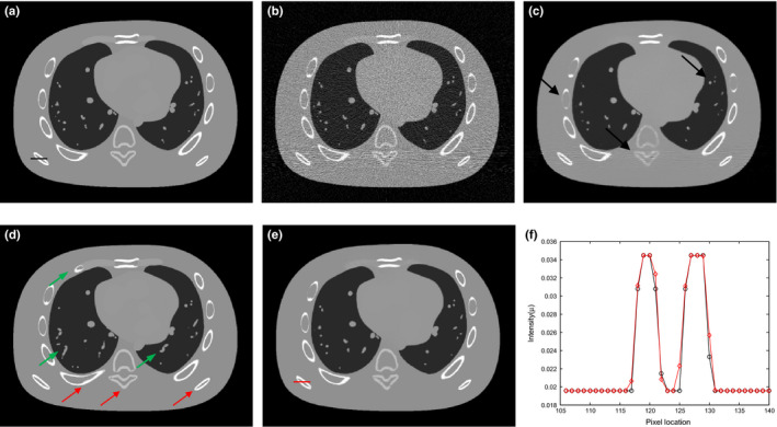
Figure 2.

Illustration of the preHQ‐guided NLM image filtering for low‐flux CT: (a) one transverse slice of the NCAT phantom; (b) FBP reconstruction from simulated noisy projection data; (c) traditional NLM filtering of low‐dose image in Fig. 2(b), SW=332, PW = 52, h = 0.015; (d) another transverse slice of the NCAT phantom serving as the prior image for preHQ‐guided NLM filtering of low‐dose image in Fig. 2(b); (e) corresponding preHQ‐guided NLM filtering result, SW = 332, PW = 52, h = 0.007; (f) profile comparison between the ground truth Fig. 2(a) and the filtering result Fig. 2(e). [Colour figure can be viewed at wileyonlinelibrary.com]
