Figure 4.

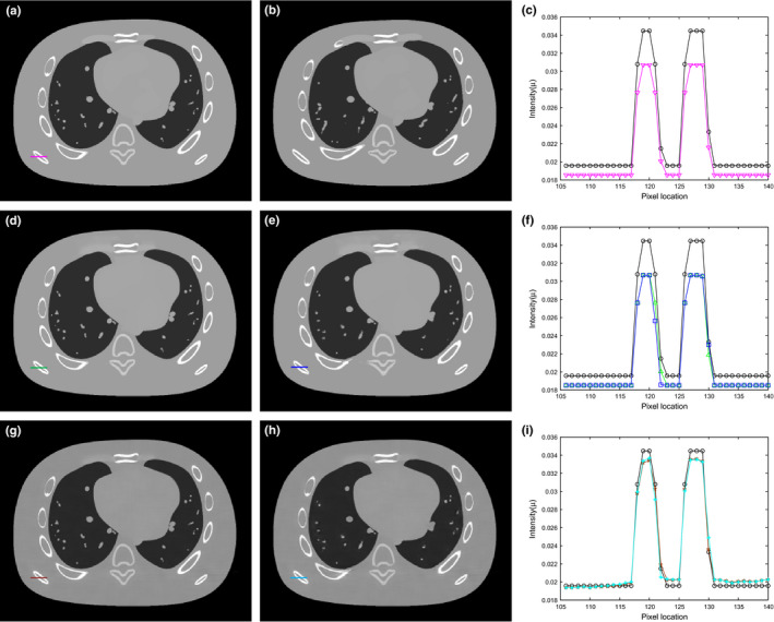
Illustration of the preHQ‐guided NLM image filtering for low‐flux CT: (a) the same transverse slice of NCAT phantom as Fig. 2(a) except for different kVp setting; (b) the same transverse slice of NCAT phantom as Fig. 2(d) except for different kVp setting; (c) profile comparison between Fig. 2(a) and Fig. 4(a); (d) preHQ‐guided NLM filtering of Fig. 2(b) using Eq. (11) with Fig. 4(a) as prior image, SW = 332, PW = 132, h = 0.007; (e) preHQ‐guided NLM filtering of Fig. 2(b) using Eq. (11) with Fig. 4(b) as prior image, SW = 332, PW = 132, h = 0.007; (f) profile comparison between Fig. 2(a) and the filtering results in Figs. 4(d) and 4e; (g) preHQ‐guided NLM filtering of Fig. 2(b) using Eq. (14) with Fig. 4(a) as prior image, SW = 332, PW=132, h = 0.007; (h) preHQ‐guided NLM filtering of Fig. 2(b) using Eq. (14) with Fig. 4(b) as prior image, SW = 332, PW = 132, h = 0.007; (i) profile comparison between Fig. 2(a) and the filtering results in Figs. 4(g) and 4(h). [Colour figure can be viewed at wileyonlinelibrary.com]
