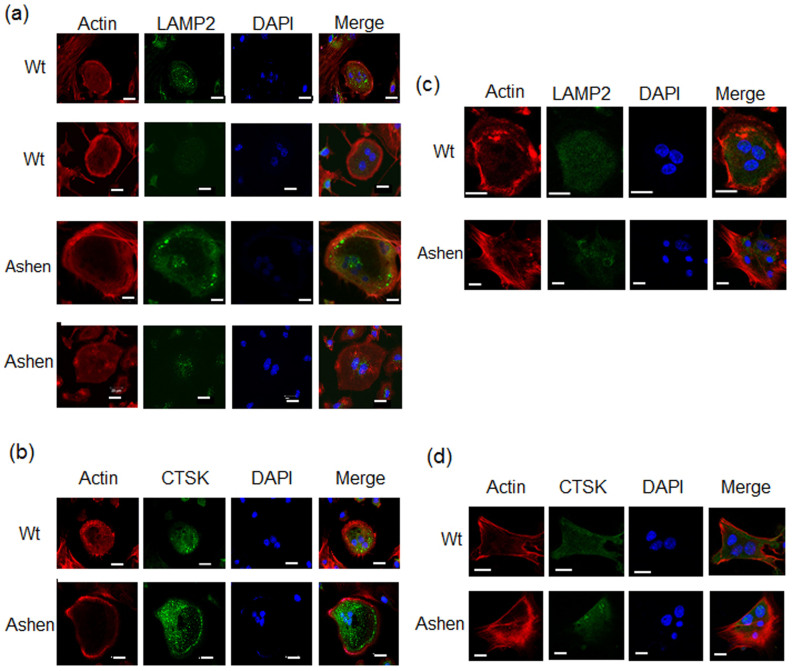
Figure 6. Immunofluorescence observation of actin ring formation and lysosomal proteins in OCLs from wild-type and ashen mice.
The cells on cover-glass were fixed, permeabilized with 0.3% Tween-20 in PBS, and then allowed to react with phalloidin for F-actin (red) or antibodies for lysosomal proteins (green). After washing, the samples were incubated with a fluorescence-labeled secondary antibody and then were visualized by confocal laser microscopy. (a) On non-coated glass slip, LAMP-2 antibody. (b) On non-coated glass slip, CTSK antibody. (c) On vitronectin-coated glass slip, LAMP-2 antibody. (d) On vitronectin-coated glass slip, CTSK antibody. Bar: 50 μm.

