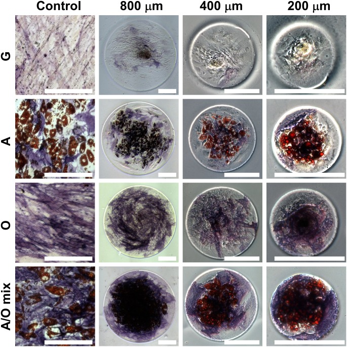
Fig 3. Differentiation of human Mesenchymal Stem Cells (hMSCs) after 15 days in culture.
Micrographs from the left to right rows show hMSCs cultivated in the control, 800, 400, and 200 mm-diameter confinements, respectively. Micrographs from upper to lower lines show hMSCs cultivated in growth, adipogenic, osteogenic, and adipogenic-osteogenic mixture media, respectively. White bar indicates 200 μm. G: Growth, A: Adipogenic, O: Osteogenic, and A/O mix: Adipogenic-osteogenic mixed media.

