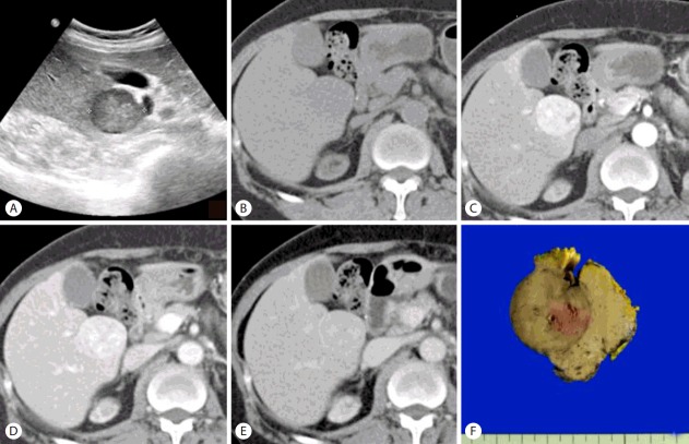
Figure 1.
Ultrasonography reveals a slightly heterogeneous hypoechoic nodule in segment 5 of the liver (S5) (A). Pre-contrast CT scan (B) shows a low-density mass of S5 of the liver with well-defined border. Contrast-enhanced CT scans show the lesion is heterogeneously and significantly enhanced on arterial phase (C), slightly hypodense on portal venous phase (D) and enhancing rim on delayed phase (E), suggestive of hepatocellular carcinoma in the background of diffuse liver disease. On section, the mass measures 3.2×3.0 cm and a relatively well-demarcated but not encapsulated and shows brown to gray color and expansile growth pattern (F).

