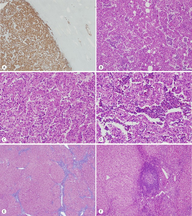
Figure 2.
The tumor was well-circumscribed along the edge of the tumor but focal foci of infiltrative growth into the surrounding non-tumorous liver parenchyme were seen in the immunostaining of HMB45 (×40) (A). The tumor mainly composed of epithelioid cells and arranged in trabecular growth pattern (×100) (B). The epithelioid tumor cells had abundant granular eosinophilic cytoplasm, distinct cell border, eccentrically located round nuclei with small nucleoli, and foci of mild to moderate nuclear atypism (×200) (C). Extramedullary hematopoiesis was recognized in the sinusoidal spaces between the tumor cells (×200) (D). The surrounding non-tumorous liver parenchyme showed chronic hepatitis with early cirrhotic change (Masson trichrome stain, E) and foci of lymphoid aggregate in some portal tracts (×100) (F).

