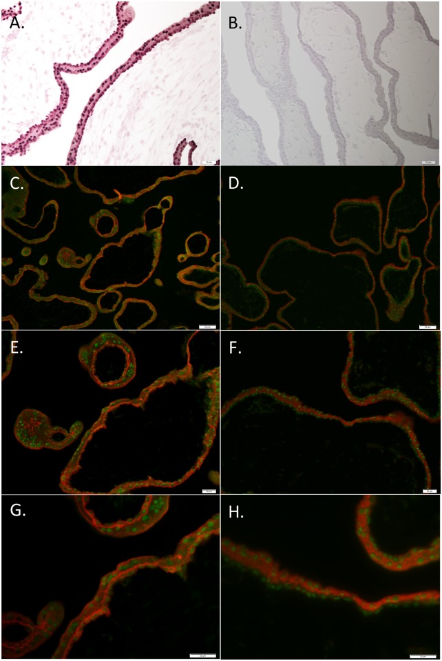
Fig 1. Immunohistochemical localization of PRR15 in 6- and 11-week human placenta.
(A) An 11-week placenta sample was stained with rabbit anti-PRR15 antibody, or (B) with the rabbit anti-PRR15 antibody that was preadsorbed with recombinant PRR15 as a negative control. Six-week placenta was used to co-localize PRR15 (green fluorescence) with KRT7 (red fluorescence; trophoblast marker; C, E, G) and with CGB (red fluorescence; syncytiotrophoblast marker; D, F, H). Collectively, these data verify that PRR15 is predominantly localized to the nucleus of both cytotrophoblasts and syncytiotrophoblasts of the human placenta. Magnification bars represent: 20 μm for A, B, E, F, G and H; 50 μm for C and D.

