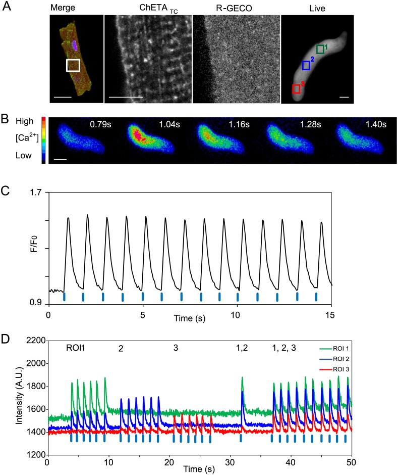
Fig 1. Expression and function of ChETATC /R-GECO in primary adult cardiomyocytes.
(A) Guinea pig ventricular cardiomyocytes were fixed and stained to reveal ChETATC (green) and R-GECO (red) two days following infection. The magnified region demonstrates regional staining differences compatible with successful 2A peptide cleavage, and membrane localisation of the optical control tool. The panel on the right shows the red fluorescence emission from R-GECO in a living cardiomyocyte; ROI’s show the stimulated regions for Fig 1D. (B) Pseudo coloured image of the single cell calcium transient revealed by R-GECO, stimulation was at t = 1sec (C). Optical stimulation with 405nm light pulses (blue tabs) every second generates synchronised calcium transients in single adult ventricular cardiomyocytes. (D) Subcellular observation and control windows in single cells. ROI’s as shown in Fig 1A were stimulated with 405nm light singly or in combination as documented in the text above the trace. Raw intensity changes for the ROI’s are shown across the experiment. Scale bar = 10μm.

