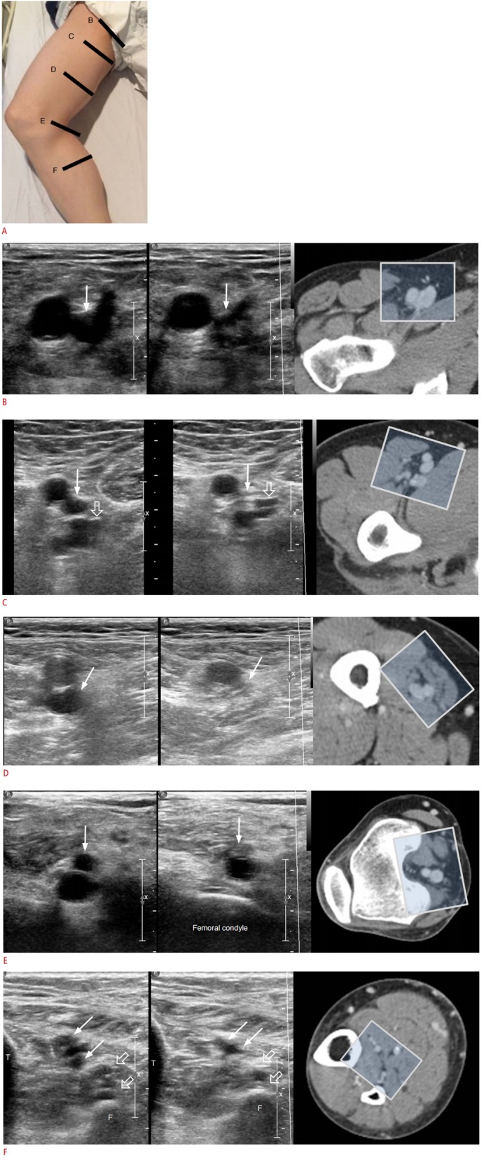
Fig. 4. Demonstration of side-by-side transverse ultrasonography (noncompression and compression views) and the corresponding sonic window in computed tomography venography based on transducer placement.

A. Patient position and schematic representation of the transducer locations are shown here (B, C, D, E, and F). B. The common femoral vein is seen at the level of the inguinal ligament on the medial side of the common femoral artery, which is round and pulsatile. The vein collapses upon compression (arrows). C. Upon following the common femoral vein downward, it bifurcates into the deep femoral vein (open arrows) and the femoral vein (arrows).
