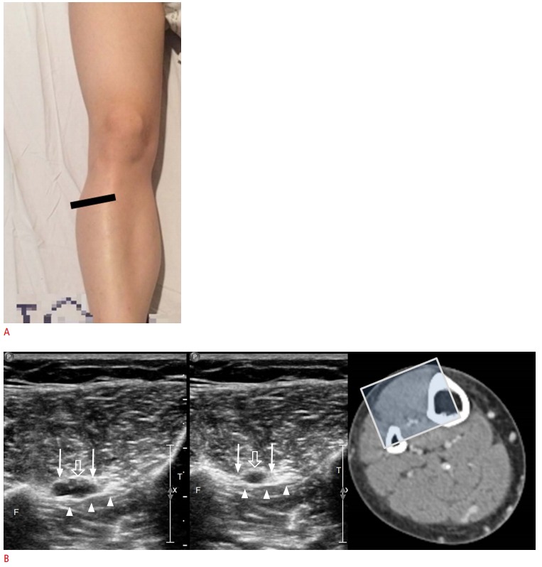
Fig. 5. Ultrasonographic findings of the anterior tibial vein.

A. Stretching of the patient’s leg permits approach to the anterior tibial vein from the anterior side. B. Above the interosseous membrane (arrowheads) between the tibia (T) and the fibula (F), the anterior tibial vein (arrows) and artery (open arrows) are visible. The sonic window is demonstrated through computed tomography venography.
