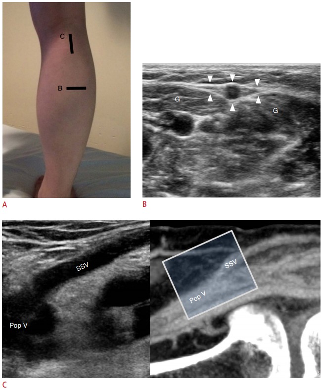
Fig. 8. Ultrasonographic findings of the small saphenous vein (SSV).

A. Patient position and schematic representation of the transducer locations are shown here. B. In the transverse view of the posterior calf, the SSV is seen in the middle of the gastrocnemius (G) belly in the fascial trunk (arrowheads). C. Longitudinal ultrasound view and the corresponding sonic window in computed tomography venography based on transducer location are demonstrated. The SSV joins the popliteal vein (Pop V) through the saphenopopliteal junction. However, many variations are noted in this region, and the saphenopopliteal junction can be absent or hypoplastic.
