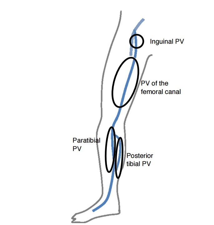
Fig. 9. Schematic images of the perforating veins (PVs) along the medial aspect of the lower extremity.

The PV of the medial leg includes the paratibial PV and the posterior tibial PV. The medial thigh perforator includes the PV of the femoral canal and the inguinal PV.
