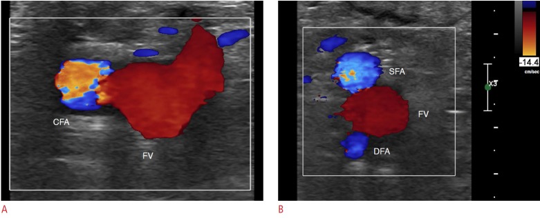
Fig. 3. Normal color Doppler ultrasonography of the femoral arteries in the inguinal area.
A. The common femoral artery (CFA) is lateral to the femoral vein (FV) on a transverse scan at the inguinal crease. Note that the size of the color box is as small as possible. B. The superficial femoral artery (SFA) and the deep femoral artery (DFA) make a shape like Mickey Mouse’s ears, and the FV forms Mickey Mouse’s face.

