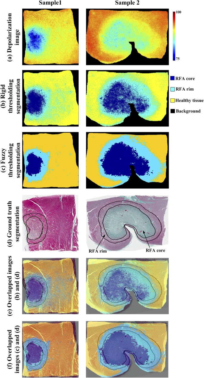
Fig 3. Representative polarization images from two ablated myocardium samples, segmented into RFA core, RFA rim, and healthy regions.
(a) depolarization images; (b) automated segmentation with global rigid thresholding; (c) automated segmentation using local fuzzy thresholding algorithm. Pseudo-colors dark blue, light blue and yellow show RFA core, rim and healthy regions, respectively. Black represents the background; (d) segmented histology image (ground truth) where demarcation of RFA core and rim regions are indicated by the black contours; (e) overlapped images from (b) and (d) showing global rigid thresholding and ground truth segmentation; (f) overlapped images from (c) and (d) demonstrating good qualitative agreement of the local fuzzy thresholding and ground truth segmentation scheme. Local fuzzy thresholding shows better qualitative agreement than the global thresholding, which overestimates the extent of the rim region. Quantitative results are summarized in Table 1 and Figs 4 and 5.

