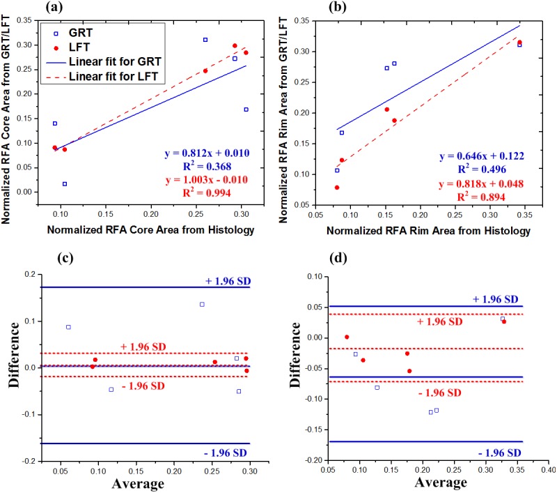
Fig 5. Comparison of both thresholding algorithms with ground truth segmentation of RF ablated tissue.

Normalized segment area calculated from global rigid thresholding (GRT, blue hollow squares) and local fuzzy thresholding (LFT, red solid circles) plotted against normalized segment area from ground truth histopathology, for (a) RFA core and (b) RFA rim region. For both RFA core and RFA rim, local fuzzy thresholding provided better correlations. Corresponding Bland–Altman plots for (c) core and (d) rim show no biases (no data points outside +/- 1.96 standard deviations).
