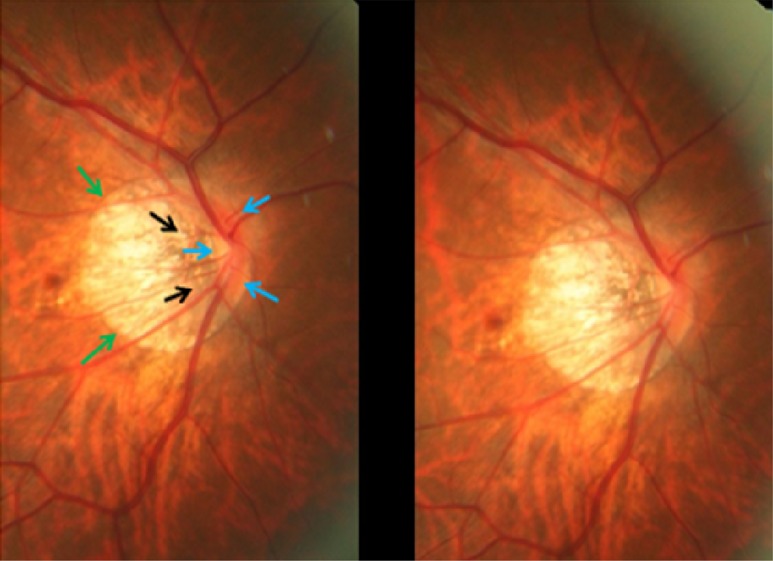
Fig 5. Stereoscopic photograph of an optic nerve head in a highly myopic eye without glaucomatous optic nerve damage, with parapapillary gamma zone (green arrows) and parapapillary delta zone (black arrows); blue arrows: optic disc border (peripapillary ring).
The neuroretinal rim at the inferior disc pole and superior pole is markedly wider than in the temporal disc region in this optic disc with pronounced rotation around the vertical axis and a slight rotation (about 15°) around the sagittal axis.

