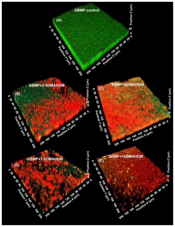
Fig. 2.
Representative CLSM image of 3D biofilms cultured for 2 days on bonding agents: (A) SBMP control, (B) SBMP+2.5DMAHDM, (C) SBMP+5DMAHDM, (D) SBMP+7.5DMAHDM, (E) SBMP+10DMAHDM. The x and x axes are parallel to the resin surface. The z axis is perpendicular to the resin surface. Live bacteria were stained green, and bacteria with compromised membranes were stained red.

