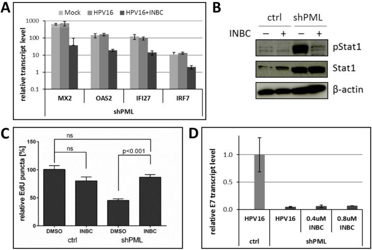
Fig 6. Inhibition of the JAK/Stat signaling pathway in PML-deficient HaCaT cells.
(A) Expression of ISGs in cells infected with HPV16 quasivirions in the presence or absence of Jak1/Jak2 kinase inhibitor. Cells were treated with 0.4 μM of INBC 1 h prior to infection. Samples were collected at 48 hpi and analyzed using RT-qPCR. The data shown are fold changes relative to mock-treated vector control HaCaT cells. Error bars represent SEM of two independent experiments. Student's t-test was used to determine P value. * P<0.01. (B) Immunoblot analysis of pStat1 and Stat1 in lysates from control and shPML HaCaT cells treated for 8 h with 0.8 μM INBC. β-actin was used as a loading control. (C) Quantification of EdU-labeled viral pseudogenome in the nuclei of cells infected with HPV16 pseudovirions in the presence of INBC. Stably transduced HaCaT cells were grown on coverslips infected with HPV16 pseudovirus in absence or presence of INBC (0.4 μM). At 30 hpi, samples were fixed and stained for EdU, PML, lamin A/C and DAPI. Number of EdU-labeled viral genome in the nuclei was counted in z-stacks spanning the whole nucleus for each cell. The data shown are average percent of viral pseudogenome present in the nuclei of vector control cells. Results were calculated in total from 60 to 116 cells collected from three independent experiments. Error bars represent the SEM, P value was determined using Student's t-test. (D) Indicated HaCaT cell derivatives were infected with HPV16 quasivirions in the absence or presence of INBC for 48 h. Relative E7 transcript levels were determined by RT-qPCR.

