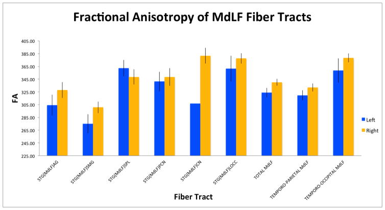
Figure 5.
Graphical representations of the fractional anisotropy (A) and volume (B) of six left and right fiber connections of the human middle longitudinal fascicle (MdLF) in 70 healthy subjects, based on the data in Table 1. Four of these connections are temporo-parietal: superior temporal-angular gyrus, STG(MdLF)AG; superior temporal-supramarginal, STG(MdLF)SMG; superior temporal-superior parietal lobule, STG(MdLF)SPL; and superior temporal-precuneus, STG(MdLF)PCN. The two temporo-occipital conections are the superior temporal-cuneus, STG(MdLF)CN, and superior temporal-lateral occipital, STG(MdLF)LOCC.


