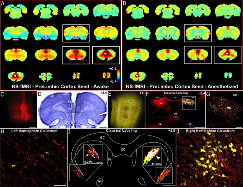
Fig. 5.

RS-fMRI in the awake state reveals functional connections of the mPFC that are lost during anesthesia. (a) Average RS-fMRI of awake rats, with a unilateral seed (white voxels) in mPFC (prelimbic cortex, PrL). Bilateral functional connectivity was observed between mPFC and the claustrum, mediodorsal thalamus (MD), and retrosplenial and entorhinal cortices. Color bar represents correlation coefficients and applies to panel b as well. (b) Anesthesia abolished functional connectivity with MD and severely reduced functional connectivity with claustrum (see pink arrows). (c–e) Anterograde tracer (Fluororuby, FR, red, left hemisphere, panel c) and retrograde tracer (Fluorogold, FG, yellow, right hemisphere, panel e) injections in PrL revealed anatomical connectivity with MD and claustrum. Scale bars: 250μm in c; 1mm in d. (f–g) Labeling was predominantly ipsilateral in the thalamus for both tracers. Anterogradely-labeled terminals were also observed in contralateral MD, terminating around retrogradely-labeled neurons, demonstrating an interhemispheric cortico-thalamo-cortical loop. Scale bars: 500μm in f; 100μm in g. Abbreviations: sm, stria medularis; 3V, third ventricle, interanteromedial nucleus, IAM; ventromedial nucleus, VM. (h–j) Anterograde projections terminated in the contralateral claustrum, intermingling with retrogradely-labeled FG neurons (panel j), demonstrating an interhemispheric cortico-claustro-cortical loop. Panel i shows a digital reconstruction of tracer labeling in claustrum and overlap analysis (50μm2 bins) in the insets. Overlapping bins (white) contained at least 4 FR-labeled terminals and 1 FG-labeled neuron. Scale bars: 50μm in h; 1mm in i, 200μm in inset of i. Abbreviations: cc, corpus callosum; ec, external capsule; lv, lateral ventricle; ac, anterior commissure.
