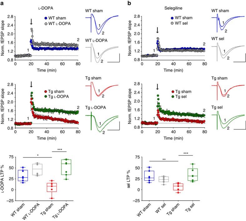
Figure 5. Sub-chronic L-DOPA or selegiline treatment rescues CA3-to-CA1 plasticity deficits in 6-month-old Tg2576 mice.
(a,b) Running plots show normalized fEPSP mean slope (±s.e.m. displayed every 2 min) recorded from the dendritic region of CA1 neurons in hippocampal slices from 6-month-old saline-treated (sham) and L-DOPA- (a) or selegiline (sel)-treated (b) WT and Tg2576 mice. The arrows indicate when a high frequency train was delivered by stimulating the Schaffer collateral pathway in the CA3 region. Traces are superimposed fEPSPs recorded during baseline (1) and 1 h after the train (2). The box-and-whisker plots below indicate the degree of potentiation, measured as fEPSP slope increase from baseline, 55–60 min after the train (a, WT: n=6 slices from 3 sham, 6 slices from 4 L-DOPA-treated mice; Tg: n=6 slices from 3 sham, 7 slices from 3 L-DOPA-treated mice; two-way ANOVA: genotype × treatment, F1,21=6.51, P=0.019; genotype, F1,21=3.22, P=0.087; treatment, F1,21=26.09, P<1.00 × 10⁻⁴; WT sham-Tg sham *P<0.050, Tg sham-Tg L-DOPA ***P<0.001 with Bonferroni's post hoc test. (b) WT: n=7 slices from 3 sham mice, 8 slices from 4 sel-treated mice; Tg2576: n=6 slices from 3 sham, 6 slices from 4 sel-treated mice; two-way ANOVA: genotype × treatment, F1,23=16.61, P=5.00 × 10⁻⁴; genotype, F1,23=2.00, P=0.171; treatment, F1,23=5.99, P=0.022; WT sham-Tg sham **P<0.010, Tg sham-Tg sel ***P<0.001 with Bonferroni's post hoc test. Both L-DOPA and selegiline increase LTP in Tg2576 mice while having no effect on WT animals (scale bars for traces: 100 μV, 10 ms).

