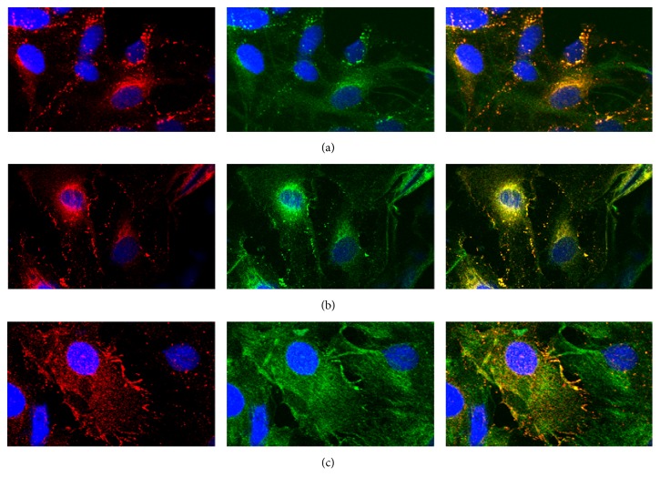
Figure 3.
Representative confocal images showed Cx43 distribution under IR/OGDR conditions at 6 h after reperfusion (red, Cx43; green, GFAP; blue, Hoechst 33342; magnification was 400x, resp.). (a) Under normal cultivation conditions, the distribution of Cx43 is similar to that in Figure 1(a). (b) Under IR/OGDR conditions with DMSO (as the medium to dissolve dynasore), the distribution of Cx43 was almost the same as that in Figure 2(b). (c) Under IR/OGDR conditions with dynasore, Cx43 was distributed in the membrane, cytoplasm, and nucleus, with the highest content in the cytoplasm. The particles in the cytoplasm of this group were significantly fewer than those of the DMSO control group (P = 0.021), and the number of particles transported into cytoplasm was reduced by 29.2%.

