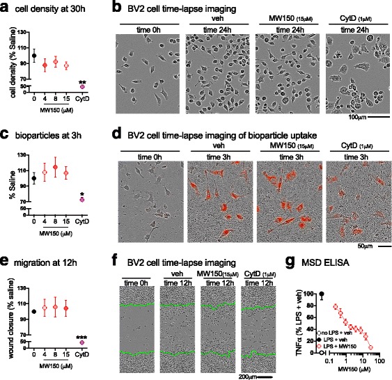
Fig. 7.

No effect of MW150 treatment on BV2 cell proliferation, migration, and phagocytosis. a Average BV2 cell density at 30 h after plating in a 96-well plate at 5000 cells/well. b A representative example of the cell density at 24 h after treatment with saline, MW150 (0, 3.75, 7.5, or 15 μM), or cytochalasin D (cytD;1 μM) (mean ± SEM, n = 3 independent experiments; 8 technical replicates included for each experiment). c Quantification of pHrodo-labeled E. coli bioparticles at 3 h after addition of bioparticles. d A representative example of the cell bioparticles uptake at 3 h after treatment with saline, MW150 (0, 3.75, 7.5, or 15 μM), or cytD (1 μM) (mean ± SEM, n = 3 independent experiments; 4 technical replicates included for each experiment). e Average size of scratch wound that is filled with cells, as determined by the percent confluency in the area left nearly devoid of cells after the scratch wound, normalized to veh at 12 h post scratch. f Representative images of the scratch wound made (highlighted by green lines), at time 0 and 12 h post scratch (mean ± SEM, n = 3 independent experiments; 8 technical replicates included for each experiment). g MW150 concentration-dependent inhibition of TNFα levels in LPS-stimulated BV2 cells (mean ± SEM, n = 1–3 independent experiments; 4 technical replicates included for each experiment). Source data is available in Additional file 4: Table S4
