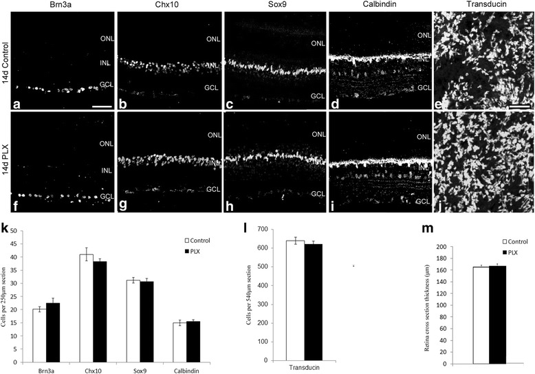
Fig. 6.

PLX ablates microglia without affecting other retinal cells. Retinal sections from mice kept on the PLX or vehicle chow diet for 14 days were labeled for ganglion cells (Brn3a; a, f), bipolar cells (Chx10; b, g), Müller glia (Sox9; c, h), and horizontal cells (Calbindin; d, i). Cell counts of images for the labeled markers (n = 8) showed no difference between the control and PLX-treated mice (k; Images taken were within 200 μm from the optic nerve). 60× images of retinal flatmounts of PLX and control-treated mice were labeled for Gα transducin (e, j). Cell counts showed no difference in the two treatments (l). Retinal thickness was also assessed in control and PLX retinas and showed no difference (m). Magnification bar in a = 50 μm, for images (a–d, f–i). Magnification bar in e = 10 μm, for images (e, j)
