Fig. 1.

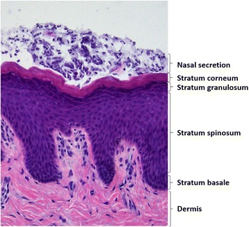
Representative histological view of epidermis in an S. aureus nasal carrier. Nasal secretion and four stratified cell layers in epidermis are marked. Sections were stained with hematoxylin-eosin, and examined in light microscope

Representative histological view of epidermis in an S. aureus nasal carrier. Nasal secretion and four stratified cell layers in epidermis are marked. Sections were stained with hematoxylin-eosin, and examined in light microscope