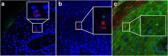
Fig. 3.

Localization of S. aureus in stratum spinosum by CLSM. Inset represents a zoomed portion of the image. Scale bar is in micrometers. We used a CLSM ×63 objective. The sections are oriented so that the outermost epidermis layer is shown in the upper left corner. a-b Green fluorescence labeling of S. aureus, blue fluorescence labeling of keratinocyte cell nuclei and S. aureus DNA. c Same labeling as in a and b, but in addition we used red fluorescence labeling of actin. Primary antibody rabbit polyclonal antibody to S. aureus (ab20920, Abcam), secondary antibody Alexa Fluor 488® goat anti-rabbit IgG (Molecular Probe™, Thermo Fisher Scientific), Alexa Fluor 594 Phalloidin (Molecular Probes™, Thermo Fisher Scientific) and DRAQ5 (BioStatus)
