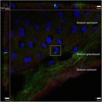
Fig. 7.

Intracellular localization of S. aureus in nasal epithelial cells. S. aureus is labeled with primary rabbit polyclonal antibody to S. aureus (Abcam), secondary antibody Alexa Fluor 488 goat anti-rabbit IgG (green) (Molecular Probes™, Thermo Fisher Scientific), DRAQ5 (BioStatus) for keratinocyte nuclei (blue) and Alexa Fluor 594 Phalloidin (A12381, Molecular Probes™; Thermo Fisher Scientific) for actin (red). Confocal laser scanning microscopy of frozen sections. Projection is constructed from confocal Z-stacks (0,2 um thick), 63× objective. Image to the left and on top corresponds to a vertical view in the z-plane. Z-plane images reveal a single cellular nucleus (blue) closely related to fluorescing S. aureus (green)
