
Figure 3.
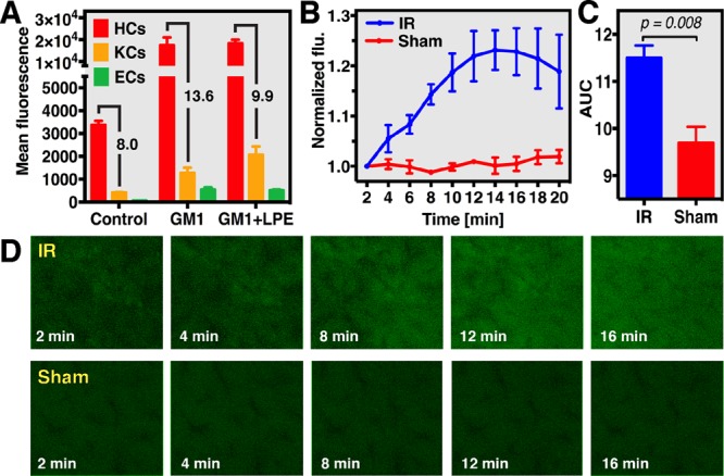
Real-time analysis of hepatocellular oxidative stress using liposome-delivered CDCFH2 during hepatic ischemia-reperfusion (IR) and sham operation in mouse livers. (A) Uptake of NBD-labeled GM1 and GM1 + lactosyl-PE (LPE) liposomes by hepatocytes (HCs), Kupffer cells (KCs), and endothelial cells (ECs), which was analyzed using flow cytometry. Oxidant formation during IR was analyzed by intravital fluorescence (flu) microscopy (D) and spectroscopy (B) in a standardized mouse model of liver IR (60 min ischemia) using CDCFH2-encapsulating GM1 liposomes. (C) Cumulative fluorescence formation during 20 min reperfusion, which was significantly higher in the IR group compared to sham controls.