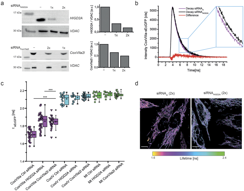
Figure 2. The lifetime of CoxVIIIa-sEcGFP increases in cells with decreased levels of scaffold proteins.
(a) Silencing of HIGD2A and CoxVIIa2l in stable CoxVIIIa-sEcGFP cells as shown by immuno-staining. Scrambled siRNA was used as a control, loading control was VDAC (middle panel). Right panel: ratio between HIGD2A respectively CoxVIIa2l and VDAC levels. (b) TCSPC diagram showing the change in fluorescence decay with decreased scaffold proteins. (c) Average fluorescence lifetimes τamp of stable HeLa cell lines expressing CoxVIIIa-sEcGFP, CoxIV-sEcGFP and mt-sEcGFP, respectively with downregulated HIGD2A or CoxVIIa2l. One data point per cell, error bars represent s.d. of ∼24 cells (n = 3 biological replicates). (d) Fluorescence intensity/lifetime images of CoxVIIIa-sEcGFP in cells with decreased scaffold protein HIGD2A or All-Stars non-targeting siRNA. Scale bars: 10 μm. Significance: ***P < 0.001 compared to CoxVIIIa-sEcGFP (ANOVA one-way).

