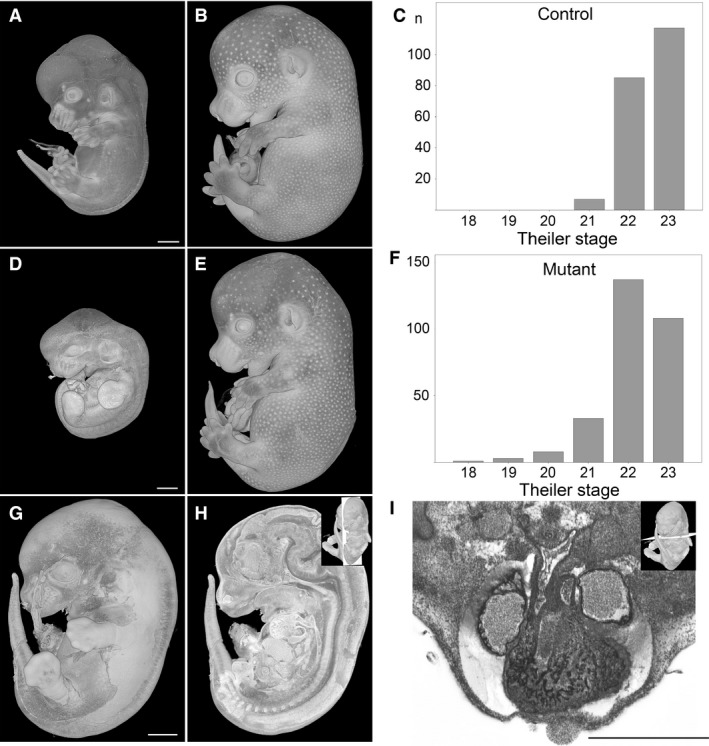
Figure 1.

Developmental progress of E14.5 embryos. (A–C) Theiler stages (TS) of control embryos range from TS21 (A) to TS23 (B). (D–F) TS of mutants range from TS18 to TS23 (E). Note that panel D shows a TS19 mutant. (G–I) TS21 mutant in which autolysis has started. Scoring of the major organs and detection of malformations such as double outlet right ventricle (I) is possible. (A,B,D,E,G) Volume‐rendered 3D models. (H) Volume‐rendered model sectioned in the median sagittal plane. (I) Axial HREM section. Scale bars: 1 mm.
