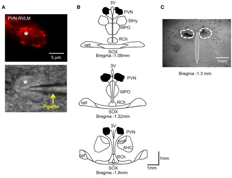
Figure 6.
(A) PVN-RVLM neuron with red fluorescence from retrograde label (top) and same neuron with patch pipette positioned on cell surface with DIC microscopy (bottom). (B) Schematic drawings of coronal sections throughout the rat hypothalamus. Shaded area indicates spread of injected dye used to mark the injection sites in the bilateral PVN. The shape of each area was determined by overlaying tracings of the outermost diffusion area of injected dye (100 nl) through the rostral-caudal plane of the PVN. (C) Representative coronal slice through the PVN demonstrating spread of injected dye. AH, anterior hypothalamic area; 3V, third cerebral ventricle; RCh, retrochiasmatic area; MPO, medial preoptic nucleus; opt, optic tract; SOX, supraopticdecussation; StHy, striohypothalamic nucleus.

