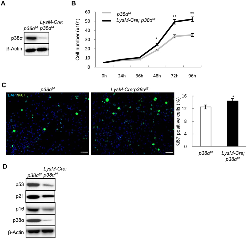
Figure 1. p38α deficiency promoted monocyte proliferation and osteoclast differentiation.
(A) Western blot results showed that p38α was deleted in the monocyte cultures of the LysM-Cre; p38αf/f mice. Bone marrow monocytes were isolated from mutant and control mice, cultured in the presence of M-CSF and RANKL for 2 days, and then used for western blot analysis. (B) p38α deficient monocytes showed increased proliferation. Bone marrow monocytes were isolated from the mutant and control mice, plated at the same number, cultured in the presence of M-CSF/RANKL, and counted at different time points. N = 3. (C) p38α deficiency increased cell proliferation in monocyte cultures. WT and p38α deficient monocytes were induced to differentiate by M-CSF/RANKL. At day 2, these cells were immunostained for Ki67 to detect S phase cells (left panel). The ratios of Ki67 positive cells to DAPI-stained cells were presented (right panel). Scale bar, 50 μm. N = 3. (D) Western blot analysis of extracts of cells described in Fig. 1A revealed that p38α deficient monocyte cultures showed a decrease in the levels of anti-proliferation proteins p53, p21, and p16. For all results in Fig. 1, P-values are based on Student’s t-test. *p < 0.05, **p < 0.01 when the value of mutant mice or cells was compared to that of control mice or cells.

