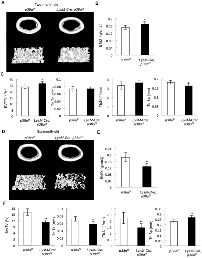
Figure 3. Micro-CT results revealed that LysM-Cre; p38αf/f mice developed osteoporotic phenotypes at 6 month of age.
(A) A representative μCT image of the femur bones of 2.5 month-old LysM-Cre; p38αf/f and control mice. (B) Two and half-month-old LysM-Cre; p38αf/f mice, compared to control mice, showed a minor increase in bone mineral density in the femur trabecular bones. N = 8. (C) Two and half-month-old LysM-Cre; p38αf/f mice only showed a minor increase in BV/TV and a decrease in trabecular separation, but not a change in trabecular number or thickness compared to control mice. N = 8. (D) A representative μCT image of the femur bones of 6 month-old LysM-Cre; p38αf/f and control mice. (E) Bone mineral density of the femur trabecular bones of 6 month-old LysM-Cre; p38αf/f and control mice. N = 8. (F) Six-month-old LysM-Cre; p38αf/f mice showed a decrease in BV/TV, trabecular number, trabecular thickness, and an increase in trabecular separation compared to control mice. N = 8. For all results in Fig. 3, P-values are based on Student’s t-test. *p < 0.05, **p < 0.01 when the value of mutant mice was compared to that of control mice.

