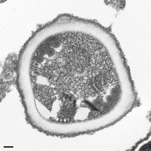
Figure 2.

Transmission electron microscopy of microsporidia identified in allograft samples from right kidney recipient. The organism shows cross-sections through the polar tube with up to 6 coils and a unikaryotic nucleus, which is characteristic of Encephalitozoon cuniculi. Scale bar indicates 100 nm.
