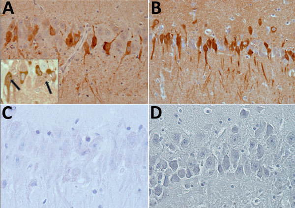
Figure 1.

Immunohistochemical detection of bornavirus X protein (A) and phosphoprotein (B) in hippocampal neurons of a brain of a Prevost’s squirrel (Callosciurus prevostii) collected in Germany in 2015. Viral antigen is shown in nuclei or cytoplasm and processes. Inset shows intranuclear dot (inclusion body) in cells with and without cytoplasmic immunostaining (arrows). No staining was observed for bornavirus X protein (C) or phosphoprotein (D) in a bornavirus-negative variegated squirrel. Original magnification ×400
