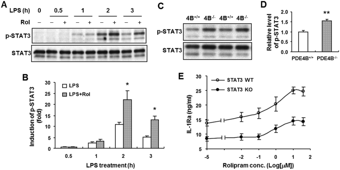
Figure 7. STAT3 phosphorylation enhanced by rolipram or PDE4B ablation does not contribute to the increased IL-1Ra production in LPS-stimulated macrophages.
Peritoneal macrophages from PDE4B+/+ mice were incubated with LPS (100 ng/ml) in the presence or absence of 10 μM rolipram for indicated times (A), or the cells from both PDE4B+/+ and PDE4B−/− mice were treated with LPS (100 ng/ml) for 2 h (C). The STAT3 phosphorylation (Tyr705) was detected by immunoblotting as described in Methods (A and C). Representative Western blots are shown. The blots were cropped for improving clarity and full-length blots are presented in Supplementary Figs 4 and 5. The level of phosphorylation relative to total STAT3 protein was quantified and expressed as fold induction to unstimulated control (B) or to the PDE4B+/+ group (D). *P < 0.05, compared with the corresponding groups treated with LPS alone (n = 4 in B); **P < 0.005, compared with the PDE4B+/+ group (n = 3 in D). (E) Bone marrow-derived macrophages prepared from STAT3+/+ and STAT3−/− mice were incubated with increasing concentrations of rolipram for 20 min prior to LPS (100 ng/ml) stimulation for 8 h. IL-1Ra accumulation in the medium was measured by ELISA. Data are the mean ± SEM (n = 7–8).

