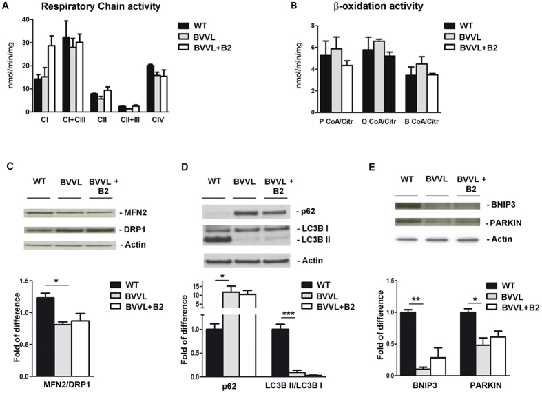
Figure 5. Mitochondrial function and autophagy in BBVL MNs before and after B2 treatment.
Respiratory chain activity (A) and β-oxidation activity (B) analysis in BVVL-MNs, BVVL-MNs+B2, and WT-MNs. No defect in respiratory chain and β-oxidation activity was observed in BVVL cells. The enzymatic activity of respiratory complexes I–IV (cI–IV) and acetyl CoA carboxylases (P CoA, P CoA, and B CoA) was determined and normalized to citrate synthase activity. Values are expressed as nmoles/min/mg protein. (C) Western blot analysis of MFN2 and DRP1 in BVVL-MNs, BVVL-MNs+B2, and WT-MNs. The MFN2/DRP1 ratio was reduced in BVVL-MNs relative to WT-MNs (*P < 0.05, ANOVA). (D) LC3BII/I and p62 expression in WT-MNs, BVVL2-MNs, and BVVL2-MNs+B2. The LC3B-II/I ratio was reduced whereas p62 expression was increased in BVVL-MNs with respect to WT (***P < 0.0001, *P < 0.05, ANOVA). After riboflavin treatment, LC3B-II/I ratio and p62 expression showed a shift towards a WT pattern. (E) Western blot analysis of Bnip3 and Parkin in WT-MNs, BVVL-MNs, and BVVL-MNs+B2 (**P < 0.001, *P < 0.05, ANOVA). The levels of both proteins were reduced in BVVL-MNs with respect to WT-MNs.

