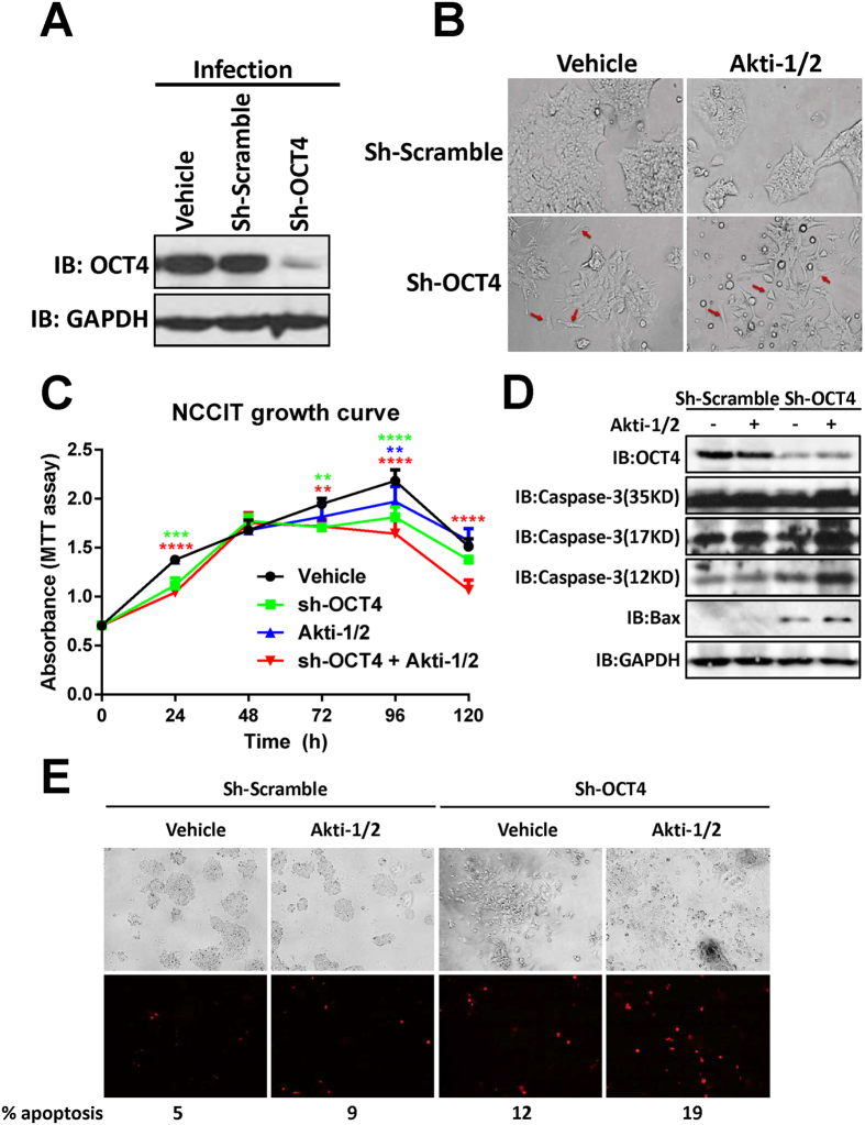
Figure 1. OCT4 shRNA + Akti-1/2 suppresses the propagation of NCCIT cells and triggers their apoptosis.
(A) NCCIT cells grown in 6-well plates were infected with culture medium only (vehicle), lentiviruses harboring scramble shRNA (sh-Scramble) or OCT4 shRNA (sh-OCT4). 3 days post infection, cells were harvested and lysed, and the whole cell lysates were subjected to immunoblotting with the indicated antibodies. (B) NCCIT cells were infected with sh-Scramble or sh-OCT4, and simultaneously treated with DMSO (vehicle) or 10 μM Akti-1/2 for 3 days. The images were captured under an Olympus IX81 microscope at 200X magnification. Red arrows indicate differentiated cells. (C) NCCIT cells grown in 96-well plates were infected with sh-OCT4 and simultaneously treated with DMSO (vehicle) or 10 μM Akti-1/2, and subjected to MTT assay at each time point. Data were expressed as mean ± SD of triplicate measurements from one of three independent experiments which gave similar results. The colored asterisks indicate the difference between each treatment group and vehicle group by significance levels (*p < 0.05, **p < 0.01, ***p < 0.001, ****p < 0.0001). (D) NCCIT cells grown in 6-well plates were treated in the same manner as (B) 3 days post infection, cells were harvested and lysed, and the whole cell lysates were subjected to immunoblotting with the indicated antibodies. (E) Adherent live NCCIT cells grown in 6-well plates were stained with 5 μg/ml Propidium Iodide for 10 min at 37 °C. Cells were examined using an Olympus fluorescence microscope at the excitation wavelength of 535 nm, the total cells and the red fluorescent cells that represent late phase apoptotic cells were counted in captured images (40X magnification), and the percentages of apoptotic cells were given at the bottom of the images.

