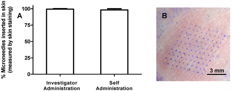
Figure 4.

Puncture efficiency of microneedle patch application as determined by the percentage of microneedles that punctured into skin. Skin sites were stained with gentian violet dye and the number of dots were counted to measure the number of microneedles that punctured into the skin. (A) Puncture efficiency of microneedle patches after investigator-administration and self-administration. Column bars represent the average percentage of microneedles that inserted into skin with standard deviation error bars shown. (B) Representative magnified image of a stained skin site showing a 10 × 10 array where the microneedles punctured into skin.
