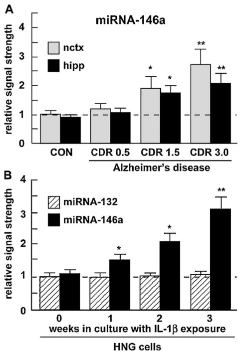
Fig. 1.
Up-regulation of an inducible miRNA-146a (A) in various stages of AD and (B) in IL-1β-stressed HNG cells. (A) Abundance of miRNA-146a in superior temporal lobe neocortex (nctx) and hippocampus (hipp) compared to 5S RNA in the same sample and to age-matched controls; number of samples used CON = 6, CDR 0.5 = 6, CDR 1.5 = 6, CDR 3.0 = 6 (3 males and 3 females in each group; see text), and (B) miRNA-146a abundance in stressed HNG cells compared to miRNA-132 in the same sample and to age-matched controls [16]. For ease of comparison control miRNA-146a levels in the neocortex arbitrarily set to 1.0; dashed horizontal line. In (B) HNG cells were treated with human recombinant IL-1β (10 ng/ml cell culture medium) and were cultured for 0–3 weeks, representing inflammatory cytokine IL-1β-mediated stress for up to one half of their in vitro lifetime of 6 weeks. The results indicate a specific up-regulation of miRNA-146a; a related miRNA-132 showed no change in either AD or in stressed HNG cells. Hence miRNA-146a is up-regulated in HNG cells stressed with inflammatory cytokines known to be abundant in AD brain. For ease of comparison control miRNA-132 levels arbitrarily set to 1.0; dashed horizontal line; N = 3–5; *p < 0.05, **p < 0.01 (ANOVA).

