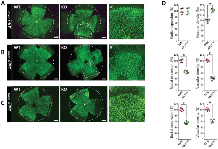
FIGURE 3. ALK2 and ALK3 promotes retinal angiogenesis.
(A–C) Representative overview of retinal vessels taken from inducible endothelial specific knock out of Alk1fl/fl;Cdh5(Pac)CreERT2 (A), Alk2fl/fl;Cdh5(Pac)CreERT2 (B), or Alk3fl/fl;Cdh5(Pac)CreERT2 (C) and their phenotypic wild-type littermates. Mice were injected with 50μg tamoxifen at P1, retinas were assayed P5; scale bar, 500μm. Both radial expansion and vascular density in the plexus region were significantly increased in P5 Alk1fl/fl;Cdh5(Pac)CreERT2 retinas (101±6.7% for radial expansion and 155±6.2% for vascular density compared to wildtype littermates). By contrast, deletion of either Alk2 or Alk3 significantly decreased both radial expansion (63.2±17.1% for Alk2fl/f;Cdh5(Pac)CreERT2 retinas and 56.4±14.2% for Alk3fl/fl;Cdh5(Pac)CreERT2 retinas compared to wildtype littermates) and vascular density in the plexus region (42.3±15.9% for Alk2fl/f;Cdh5(Pac)CreERT2 l retinas, and 61.9±12.8% for Alk3fl/fl;Cdh5(Pac)CreERT2 retinas compared to wildtype littermates). Areas within the white rectangle in middle column are shown in higher magnification (right column). While radial expansion was similarly affected by deletion of either Alk2 or Alk3, vascular density in the plexus region is more severely affected by the deletion of Alk2 than Alk3. Endothelial cells are visualized by anti- Isolectin B4 (IB4) staining. (D) Quantification of radial expansion and vascular density (% vascularized area) in inducible endothelial specific knockout of each BMPR1 (pink) and their littermates (gray) (n=5). P<0.05. Statistical significance was assessed using a Student’s unpaired t-test.

