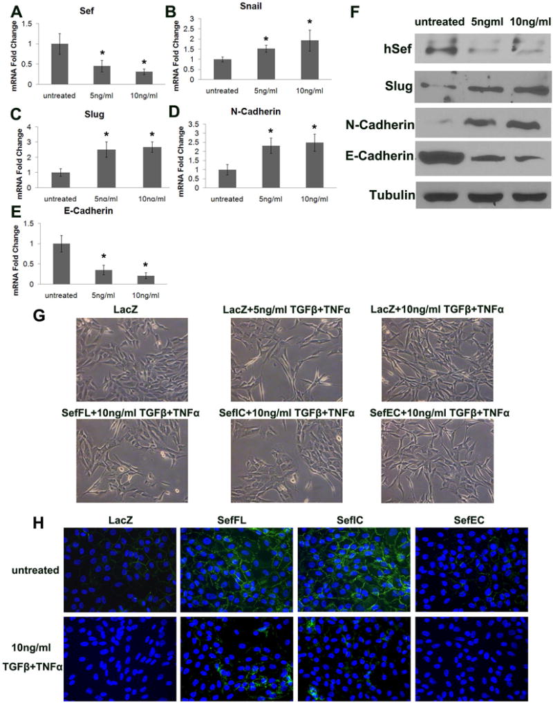
Figure 4. Sef inhibits TGFβ/TNFα induced EMT in MCF-10A cells.

(A–E) RT-qPCR analysis of Sef and EMT marker expression in MCF-10A cells treated with 2.5ng/ml TGFβ plus 2.5 ng/ml TNFα, or 5 ng/ml TGFβ plus 5 ng/ml TNFα for 48h. (A) Sef, (B) Snail, (C) Slug, (D) N-cadherin, (E) E-cadherin (* indicates p<0.05 between NT and Sef shRNA transduced cells) (F) Immunoblot analysis of Sef and EMT markers of TGFβ/TNFα treated MCF-10A cells. (G) Morphologic changes by TGFβ/TNFα treatment and AdSefFL, AdSefIC, AdSefEC overexpression. (H) E-cadherin immunostaining (green) of TGF/TNFα treated and AdSefFL, AdSefIC, AdSefEC overexpression in MCF-10A cells. Cells were counterstained with DAPI (blue).
