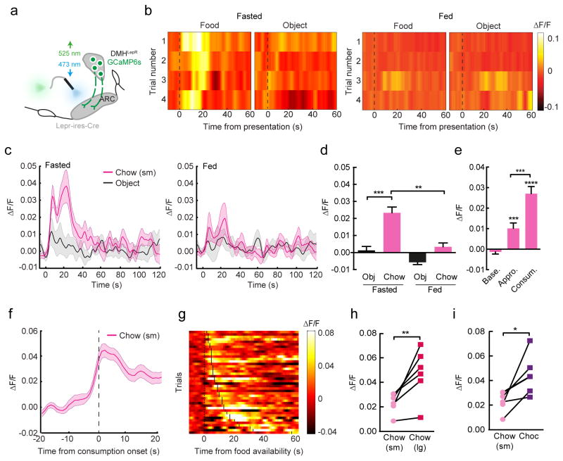
Figure 4. DMHLepR→ARC axons are rapidly activated upon sensory detection of food.
(a), the real-time activity of DMHLepR→ARC axons was determined using in vivo fiber photometry. (b–d), DMHLepR→ARC axons were rapidly activated upon presentation of a small chow pellet (t=0), compared to a non-food object, in a energy-state dependent manner (b, individual trials in one representative mouse on one day in the calorie restricted and ad libitum fed state; c, mean effects from all mice across time, n=6; d, mean response from 0–10s post food presentation, repeated measures ANOVA, F(5,15)=36.08, p<0.0001). (e), activation of DMHLepR→ARC axons to small pellet availability occurred prior to the initiation of consumption (n=36; repeated measures ANOVA, F(35,70)=35.30, p<0.0001). (f–g), Mean responses to small pellet presentation aligned to onset of consumption (f) and individual trial responses aligned to food availability (g; onset of consumption denoted with vertical black bar on each trial) demonstrating activity rising prior to consumption. (h–i), calcium response of DMHLepR→ARC axons to large chow pellets (h; n=6; paired t-test, t(6)=3.61, p=0.015) and chocolate (i; n=6; paired t-test, t(6)=3.13, p=0.026) were potentiated compared to that elicited by small chow pellets in the same mouse. All data presented as mean±SEM; post-hoc p-values *p<0.05; **p<0.01; ***p<0.001; ****P<0.0001. Abbreviations, ΔF/F, fractional change in fluorescence.

