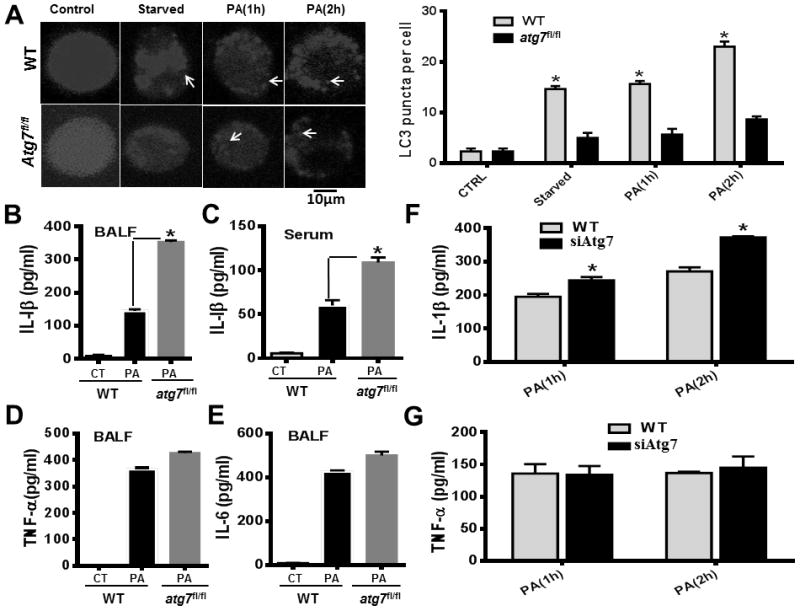
Figure 3.

Hampered autophagosome formation and increased IL-1β in Atg7-deficient macrophages and septic atg7fl/fl mice (A) AM cells were transfected with RFP-LC3 plasmid for 24 hours. Then the cells were infected with PAO1 at MOI of 10:1 for the indicated time periods. The number of LC3 puncta (arrows) within each cell was determined by confocal laser scanning microscopy. Arrows show RFP-LC3 punctation. WT mice and atg7fl/fl mice (*P<0.05; n=5, T tests.) were infected with 5×10⁶ CFU of PAO1 for 24 hours. (B) IL-1β (eBioscience, Cat #, 88-7013) production in BALF of WT mice and atg7fl/fl mice. (C) IL-1β levels in blood of WT mice and atg7fl/fl mice. (D) TNF-α (88-7324) concentrations in BALF of WT mice and atg7fl/fl mice. (E) IL-6 (88-7064) levels in BALF of WT mice and atg7fl/fl mice. (*P<0.05; n=5, T tests.). (F) IL-1β (88-7013) concentrations in siRNA-transfected and WT MH-S cells at different times. (G) TNF-α (88-7324) concentrations in siRNA-transfected and WT MH-S cells at different times. Cytokines were measured by ELISA. Data are representative of three independent experiments (*P<0.05; n=5, T tests.)
