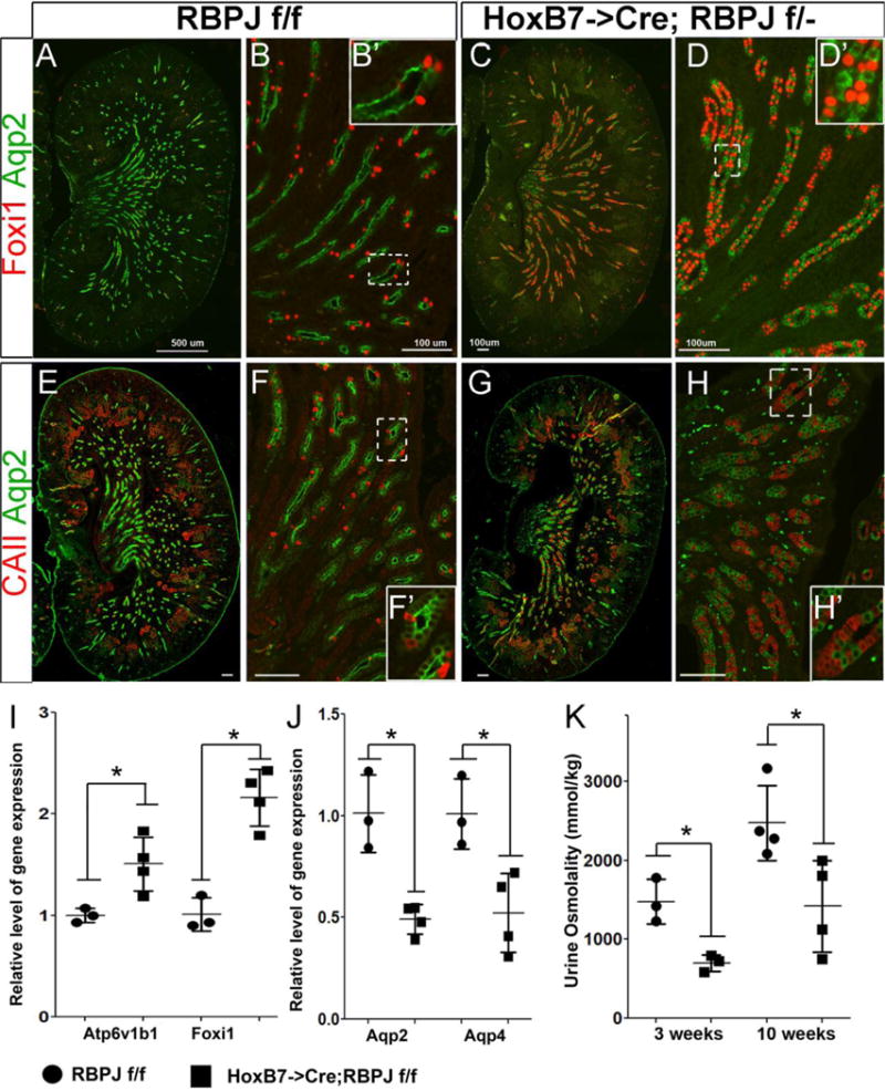
Figure 2. Notch signaling mediated patterning of collecting duct cell fates is dependent on RBPJ. A, B, E and F.

Kidney sections of wild-type post-natal day 1 RBPJf/f mice reveal many Aqp2 expressing cells intermingled with a few ICs expressing Foxi1 (A&B) or CAII (E&F) within the CDs. C, D, G and H: Kidney sections with RBPJ-deficient CDs contain many more ICs expressing Foxi1 (C&D) and CAII (G &H) and fewer Aqp2-expressing PCs. A, E, C and G are images of whole kidney sections revealing the central papillary and inner medullary regions. B, D, F and H are higher magnifications of inner medullary/papillary regions. D′ and H′ are higher magnifications of boxed areas in D and H, respectively. I &J: RT-qPCR reveals significantly increased expression of Atp6v1b1 and Foxi1, along with significantly decreased levels of Aqp2 and Aqp4 in E18.5 HoxB7->Cre;RBPJf/f kidneys (n=4) compared with RBPJf/f littermate kidneys (n=3). K. The urine concentrating ability of HoxB7->Cre;RBPJf/f mice (n=3 or 4 per time point) is significantly decreased compared with RBPJf/f wild-type littermate (n=3 or 4 per time point). A separate cohort was analyzed at each time point. * denotes p<0.05, two-tailed unpaired t-tests. Scale bars are 100μm, expect A it is 500μm.
