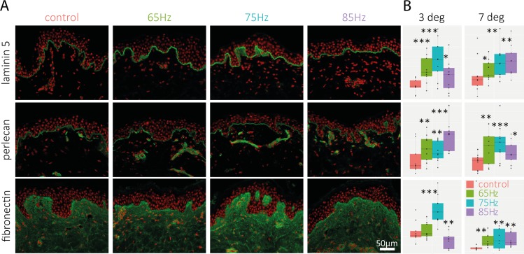
Fig 4. Immuno-labeling of DEJ and Dermal proteins for 65-85Hz treatment.
(A) Immuno-labeling of laminin-5, perlecan and fibronectin (green labeling) at D5 for three tested frequencies with a 3° peak-to-peak angular amplitude; cell nuclei are stained with a propidium iodide solution (red labeling). Data shown are collected from one 33 year old donor, after 5 days of massaging. A second 32 year old donor was analyzed with similar results (data not shown). (B)Box Plot representation of the fluorescence intensity (arbitrary scale) of the measured markers for each condition tested at D5. Data shown are collected from two donors: 3 degree data were collected from a 33year old donor, and 7deg data were collected from a 32year old donor. The stars indicate the statistical significance of the labeling quantification for each condition compared with untreated skin (***: p<0.001, **: p<0.01 *: p<0.05).

