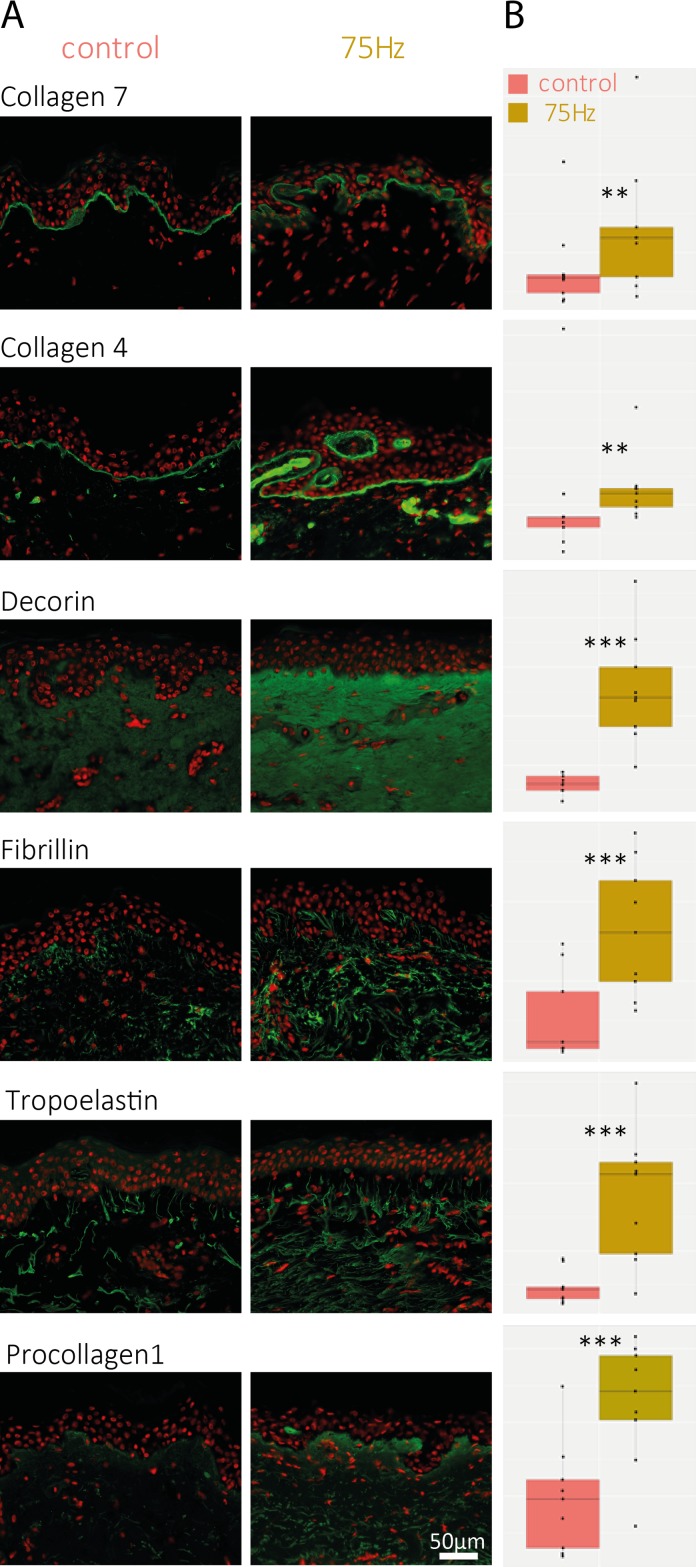
Fig 5. Immuno-labeling of DEJ and Dermal proteins for 75Hz treatment.
(A) Immuno-labeling of some dermal markers (green labeling); (B)Box Plot representation of the fluorescence intensity (arbitrary scale) of the measured markers. Data shown are collected from one 68 year old donor, after 5 days of massaging for all markers, with the exception of type VII collagen and procollagen 1, which were sampled after 10 days of treatment from the same donor. A second 50 year old donor was analyzed with similar results (data not shown). The stars indicate the statistical significance of the labeling quantification for each condition compared to untreated/control skin (***: p<0.001, **: p<0.01 *: p<0.05).

