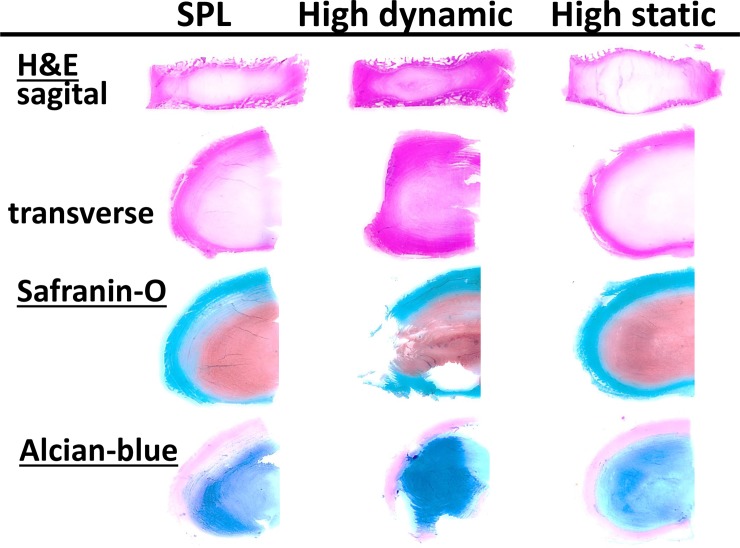
Fig 8. Histological scoring of IVD degeneration.
This overview shows representative images of histological sections from the three experimental groups after the 14-day culture and loading experiment as used for the scoring of IVD degeneration according to the Rutges scale. From top to bottom, section are: midsagittal H&E stained sections to score endplate damage (left posterior, right anterior); transverse H&E stained sections to score annulus and nucleus matrix morphology (all left haf IVDs: above anterior; left lateral; bottom posterior); transverse sections stained with Safranin-O and Alcian-blue to observe changes in proteoglycan and GAG distribution. All sections and stainings combined depict the characteristics of the transition zone. For the specific scores of each experimental group per section and a description of the observed degenerative changes due to culture and loading, please see Table 2 and the related results paragraph.

