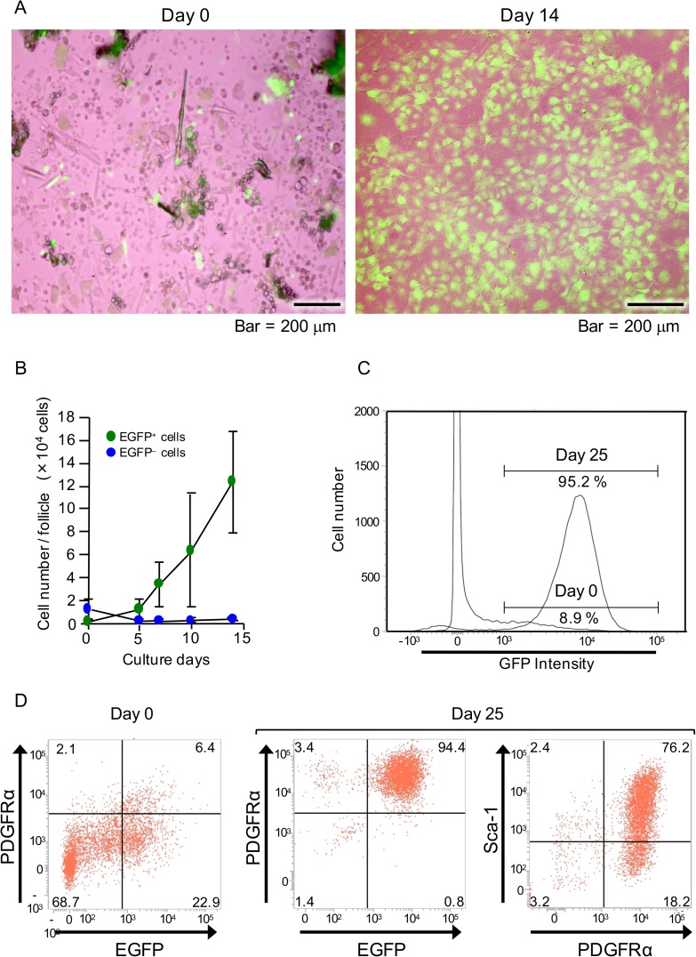
Fig 2. Examination of proliferative potential and level of mesenchymal stem cells in NCDFCs.
(A) Phase-contrast images of whisker follicle cells subsequent to enzymatic processing on day 0 (left panel) and cells cultured in stem cell growth medium for 14 days (right panel). (B) The number of EGFP+ cells per whisker follicle increased with culture time. (C) EGFP-gated flow cytometry analysis charts of whisker follicle cells. EGFP+ cells were detected immediately after sampling (day 0) as well as after 25 days of proliferation in stem cell growth medium. (D) Flow cytometry analysis of cell-surface markers using freshly isolated whisker follicle cells from P0-Cre; CAG-CAT-EGFP Tg mice (day 0) and after 25 days of proliferation in stem cell growth medium. Representative flow cytometry images revealing EGFP- and PDGFRα-gated (left and middle panels) cells, and PDGFRα- and Sca-1-gated cells (right panel). Data shown represent mean values from 3 independent experiments, with error bars indicating SD.

