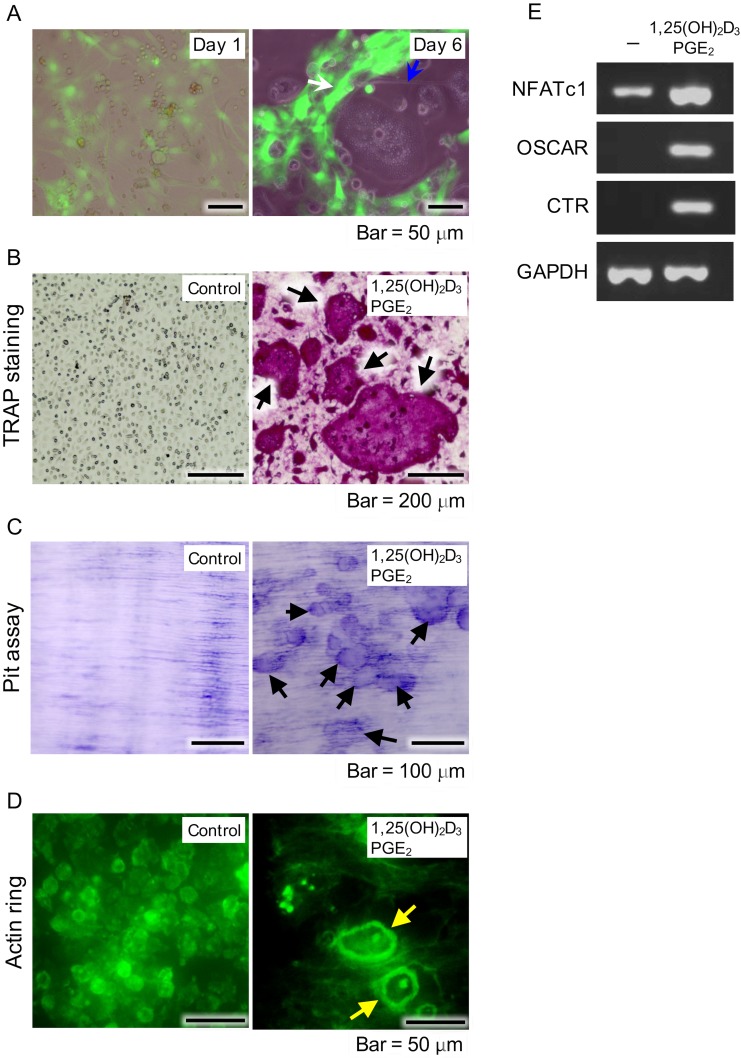
Fig 5. Examination of osteoblast functions supporting differentiation of osteoclasts.
(A) NCDFCs co-cultured with mouse bone-marrow cells including osteoclast precursor cells. Phase-contrast images of proliferative NCDFCs and mouse bone marrow cells in the presence of PGE2 and 1,25(OH)2D3 on day 1 (left panel). Formation of osteoclast-like cells (blue arrow in right panel) alongside EGFP+ cells (white arrow in right panel) on day 6. (B) Formation of osteoclasts in presence of PGE2 and 1,25(OH)2D3 detected by TRAP staining (arrows) after 6 days. (C) Cells showed bone resorption capacity after 6 days, as detected by toluidine blue staining (arrows). (D) Cells formed actin rings specific to the cytoskeleton of osteoclasts (yellow arrows), as detected by FITC-phalloidin staining after 6 days. (E) Semi quantitative RT-PCR measurements of expression of the osteoclast-related genes NFATc1, OSCAR, and calcitonin receptor in the presence of 1,25(OH)2D3 and PGE2. PGE2, prostaglandin E2; RT-PCR, reverse transcription-polymerase chain reaction.

